
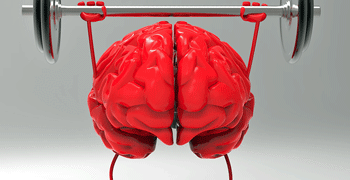


The Hanson group strikes again! Maureen Hanson is clearly determined to get at what happens during exercise in ME/CFS. Not only does she regularly employ exercise stressors (wake up, long-COVID researchers!) but in the “Temporal Dynamics of the Plasma Proteomic Landscape Reveals Maladaptation in ME/CFS Following Exertion” study, she determined what happened after not one, but two maximal exercise tests.
Hanson is throwing everything she can at ME/CFS and we’re lucky to have her, the lead researcher Arnaud Germain on this study, and her team. With their use of exercise studies to probe and molecularly profile people with ME/CFS, they’re miles ahead of most long-COVID researchers.
The Hanson group is basically trying to understand what breaks when ME/CFS patients’ systems are stressed. Proteins are a good way to assess this because these complex molecules carry out the work of the cell; i.e., protein analyses can tell us how cellular functioning has gone awry.

Proteins are complex molecules that do the work of the cell. They were the focus of this study.
Proteomics can’t tell us everything, though. Protein analyses can give us a general idea which tissue (immune, muscle, blood vessels) is not functioning well, but they lack specificity. It also cannot tell us what role the protein is playing; i.e. it can’t tell us if the change in a protein’s level is causing a problem or is the consequence of the problem.

Protein analyses are very good, though, at identifying which biological “programs” are failing. They’re also very good at tracking when the biological programs fail (before or after exercise) and if that failure results in increased symptoms.
The Hanson group used SomaScan to identify approximately 6,000 proteins in a single drop of blood. This large survey covered proteins associated with almost every major biological pathway in the human body. They then used machine learning to identify which protein levels had changed and which proteins were most effective at distinguishing ME/CFS patients.
It was a nice, large study containing 132 people with ME/CFS and sedentary, age and BMI-matched healthy controls.
THE GIST
-

Next up – find out what starts it all off. What is the cause and what is the consequence?
The Hanson group strikes again! In this study, Maureen Hanson’s group at Cornell determined what happened to protein levels after not one but two exercise tests. Proteins are important because they do the work of the cell. Proteomic studies are very good at identifying which biological “programs” may be failing.
- No change in single proteins were seen at baseline but when protein pathways were assessed, dysregulated pathways popped out in spades.
- Chief among them were pathways associated with receptors on T cells. Because T-cells interact with the body through their receptor, the widespread reduction of T-cell receptors suggested that ME/CFS patients’ T-cells were hunkered down and listless. Perhaps because they were exhausted, they’d made it difficult for the body to interact with them.
- Another theme popped up: a failure to respond. Exercise altered three times more proteins (15% of proteins) in healthy but sedentary controls than in people with ME/CFS (5%).
- A cluster analysis suggested that exercise immediately affected innate immune (i.e., inflammation) and neuromuscular signaling in ME/CFS. Exercise appears to produce a hearty dose of inflammation (complement-neutrophil activation) and oxidative stress, which plugs up the small blood vessels and reduces blood flows to the muscles.
- This is how you produce PEM without muscle damage. The muscles are not damaged – they’re just not functioning. The problem appears to be more with the blood flows to the muscles and muscle repair.
- Indeed, the peak disruption caused by the exercise occurred 24 hours after it. Instead of exercise-induced muscle damage, the main problem appears to be the inability to perform the normal muscle repair work required after exertion. (Exertion always requires muscle repair.)
- During this period of peak disruption, it appears that the body has trouble calming down the immune activation. Neuroimmune problems were also seen.
- Studies like this suggest that ME/CFS appears to be just what patients say it is: it’s a disease that causes the body to respond abnormally to stress. ME/CFS is not like cancer or diabetes or multiple sclerosis. All these diseases are wholly present at baseline (at rest), but ME/CFS only really reveals itself when the body’s systems are put under stress. That’s why it’s one of the most functionally disabling diseases known.
- This study validated numerous prior findings. Now that the main factors present in this are becoming clearer, the next steps are to develop a clear causal chain. What starts this disease off? Which factors are driving it and which are simply the consequence of it?
Results
No change in single protein levels were seen at baseline. Look at what happened, though, when groups of functionally related proteins were assessed during a GSEA (gene set enrichment analysis). Even at baseline, dysregulated protein pathways popped out in spades. We’ve seen this happen before. Pathway analyses provide much more information than analyses of individual factors.
T-Cells Pop Out
The authors reported that the GSEA revealed “compelling differences in intercellular signaling in ME/CFS cases, most notably in “T-cell receptor signaling” and other mechanisms of cell-cell communication. This was the only pathway that was dysregulated at baseline, and right after, and then 24 hours after exercise.
It was not one or two aspects of T-cell receptor signaling either. Note that almost every plot in the TCR table is orange – indicating that signaling was reduced across the board in these cells.

Check out how many orange circles there were. They indicated that T-cell receptor proteins were decreased across the board.
The fact that this dysregulation started at baseline and was maintained after exercise suggests that the T-cell signaling “set point” (i.e., the point at which the T-cells respond) was off from the beginning, and only got worse after exertion.
The upregulation of the exception – glycolysis – was noteworthy, indeed. T-cells use glycolysis to produce energy. The upregulation of this receptor suggested that they were seeking ways to increase energy production.
Ultimately, the finding – low levels of the receptors on T-cells – suggest that, even at rest, T- (and B) cells appear to be in a kind of listless state – possibly because they’re exhausted. This appears to fit perfectly with the last study Health Rising reported on and other studies which have found both T and B-cell exhaustion. Of course, this is what we want – we want findings that pop up again and again in different studies – and this study was full of them.
Long COVID – the Inflamed and Exhausted Disease? Plus, the JAK Drug Trial Boom
A Failure to Respond to Exercise…Again

Only a third as many proteins responded to exercise in ME/CFS patients as in healthy controls.
Another theme popped up: a failure to respond. Exercise only made things worse. The fact that every pathway was downregulated except for one, once again suggested that, at the molecular level, ME/CFS patients’ bodies are failing to respond to exercise.
Exercise altered three times more proteins (15% of the proteins) in healthy but sedentary controls compared to the people with ME/CFS (5%). To my mind, this finding demolishes the deconditioning hypothesis. If people with ME/CFS were deconditioned but otherwise healthy, exercise should trigger greater protein activation as the body attempts to deal with the shock it has just received. Instead, this study suggested that ME/CFS patients’ systems are simply offline.
Immediate Exercise Hit
A cluster analysis suggested that exercise immediately affected innate immune (i.e., inflammation) and neuromuscular signaling in ME/CFS; i.e., the inability of the system to respond normally begins immediately after (or during) exercise.
It appears to begin with a hearty dose of inflammation (complement-neutrophil activation) and oxidative stress, which activates the endothelial cells in the blood vessels. The oxidative stress findings were consistent with those reported in Lipkin’s recent study, which found increased oxidative stress and immune activation.
“Screaming Immune Activation”: Major Lipkin Study Finds ME/CFS Systems Folding Under the Stress
The inflammation potentially plugs up the small blood vessels and reduces blood flows to the muscles. Indeed, Wust’s deep dive into the muscles suggests that the small blood vessels’ access to the muscles is being blocked. Lacking their preferred resources (oxygen and other nutrients), the muscles respond by increasing anaerobic energy production.
Are Barriers to Blood Diffusion Causing ME/CFS and Long COVID? The 2025 Charité International Conference Pt I
This is how you produce PEM without muscle damage. The muscles are not damaged – they’re just not functioning. The problem is more with the blood flows to the muscles.
Plus, “highly significant” alterations in the “mTOR signaling pathway” suggested that exercise had impacted cellular stress, energy production, and immune activation.
(Note that “signaling” seems to be most affected. Again, this is likely good news as signaling should be easier to correct than overt damage.)
Peak Problems
Interestingly, the peak disruption produced by exercise occurred 24 hours after the first exercise stressor and impacted many pathways (15 significantly altered neuroimmune, metabolic, and inflammatory pathways).
The problem appears to be a faulty muscle repair response. This is why the most impairments showed up 24 hours after exercise – not immediately after exercise. This means that ME/CFS patients entered their next CPET exercise session with muscles that weren’t fully repaired after the first session. That’s presumably why energy production drops at the second exercise session in the 2-day exercise tests.
Three immune-related pathways, “T cell receptor signaling”, “B cell receptor signaling”, and “IL-17 signaling”, were all significantly downregulated. Note the emphasis on adaptive immune system (T and B-cells) suppression and “signaling”. The authors believe these findings reflect an inability to resolve immune activation – a theme we’re seeing more and more.
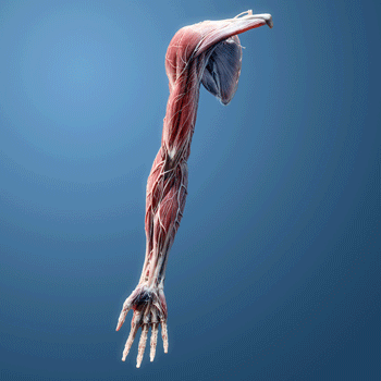
Exertion did not appear to be damaging the muscles; instead, muscle repair processes don’t appear to be kicking in.
Reductions in three muscle proteins associated with muscle structure suggested that muscle repair was impaired. Although intense exercise breaks down muscle, it ultimately builds muscle as it is remodeled post-exercise. This is not the first study to suggest that the muscle repair process is most affected in ME/CFS.
Interestingly, none of these proteins suggested the short but intense exercise session had caused muscle damage! The fact that the muscles are not actually being damaged would only seem to be good news.
It seems that any study is going to uncover nervous system issues, and this one did not disappoint. Interestingly, given the TRPM3 findings from Australia, a connection among TRP (transient receptor potential) channels emerged. So did problems with “dopaminergic synapses”.
Conclusion
From the immune system to energy production and metabolism to the nervous system, this proteome study validated what we’ve learned about ME/CFS. It’s encouraging to see similar findings pop up in studies from different research groups that often employ different means.
Studies like this suggest that ME/CFS appears to be just what patients say it is: it’s a disease that causes the body to respond abnormally to stress. This is an important distinction. ME/CFS is not like cancer or diabetes or multiple sclerosis. All these diseases are fully present at baseline (at rest), but ME/CFS only really reveals itself when the body’s systems are put under stress. That why it’s one of the most functionally disabling diseases known.
That’s why it makes sense that signaling is popping up so much. The signals that are supposed to kick in when the body is stressed aren’t.
This study did a lot, but it can’t determine causality; i.e., it can’t tell which was the first domino to fall. Given the results of other studies, it appears likely that stronger-than-normal levels of inflammation played a role. But exactly which cells are producing the inflammation, what is causing the immune activation (bits of viruses, epigenetic shifts?), and what the inflammation is affecting are not clear.

Next up – finding out what starts it all off and what is causing what.
We have lots of abnormalities (a good thing). What we now need is a clear causal chain. Are T and B-cell problems driving the problems in ME/CFS or do they result from them? Long-COVID immunomodulator and antiviral trials should prove invaluable in helping determine what’s driving what in the immune system.
Can turning the JAK/STAT system down, for instance, turn down the immune response, reduce inflammation and oxidative stress, and allow the mitochondria to produce normal amounts of ATP? By reducing inflammation, could these or other drugs heal the brain/blood barrier and normalize brain functioning?
Maybe the most important thing this and similar studies are doing is showing that core problems exist at the molecular level, and that the more researchers dig into the molecular foundations of this disease, the more they will uncover.
Donation Drive Update
Thanks to everyone who has brought us over 50% of the way to our goal!
Integration and moving forward were the themes of this blog. Look at all the links to past blogs that were posted. (We could have posted more but it would have been hard to read the actual blog.) Those links underscore what a rich resource Health Rising is. Because we’ve been digging into the nitty gritty of ME/CFS for over ten years now, we can show how X finding builds on Y finding, which built on Z finding. This field is moving forward, and we’re moving forward with it, and we’re looking forward to what’s next. If that supports you, please support us.
HEALTH RISING IS NOT A 501 (c) 3 NON-PROFIT

